You are here
Eye Anatomy
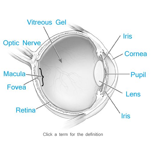
Cornea

The transparent layer forming the front of the eye that covers the iris, pupil, and anterior chamber and provides most of an eye's optical power.
Fovea
Central pit in the macula that produces sharpest vision. Contains a high concentration of cones and no retinal blood vessels.
Iris
Pigmented tissue lying behind the cornea that gives color to the eye (e.g. blue eyes) and controls amount of light entering the eye by varying the size of the pupillary opening. Most forward extension of the middle (uveal) layer of the eye; seperates the anterior chamber from posterior chamber.
Lens
Natural lens of the eye. Transparent, biconvex intraocular tissue that helps bring rays of light to focus on the retina. Suspended by fine ligaments (zonules) attached between ciliary processes.
Macula
Central area of the retina surrounding the fovea; area of acute central vision.
Optic Nerve
Second cranial nerve. Largest sensory nerve of the eye; carries impulses for sight from the retina to the brain. Composed of retinal nerve fibers that exit the eyeball through the optic disc, traverse the orbit, pass through the optic foramen into the cranial cavity, where they meet fibers from the other optic nerve at the optic chiasm.
Pupil
Variable-sized black circular opening in the center of the iris that regulates the amount of light that enters the eye.
Retina
Light sensitive nerve tissue in the eye that converts images from the eye's optical system into electrical impulses that are sent along the optc nerve to the brain, to interpret as vision. Forms a tin membranous lining of the rear two-thirds of the globe; consists of layers that include rods and cones; bipolar, amacrine, ganglion, horizontal and Muller cells; and all interconnection nerve fibers.
Vitreous Gel
Transparent, colorless gelatinous mass that fills the rear two-thirds of the eyeball, between the lens and the retina.
Home
......................................
Locations
......................................
Schedule Appointment
About
......................................
Press & Events
......................................
Testimonials
Retina Conditions
......................................
AMD - Macular Degeneration
......................................
Diabetic Eye Disease
......................................
Epiretinal Membrane
......................................
Macular Hole
......................................
Retinal Detachment
Research
......................................
Tools & Resources
......................................
Contact


