You are here
Epiretinal Membrane (Macular Pucker)
What is an Epiretinal Membrane?
The retina is a layer of light-sensing cells lining the back of your eye. As light rays enter your eye, the retina converts the rays into signals, which are sent through the optic nerve to your brain where they are recognized as images. The macula is the central portion of the retina responsible for clear, detailed vision which is needed for activities such as reading and driving. Damage to your macula causes blurred central vision, making it difficult to perform tasks such as reading small print, threading a needle, or reading a street sign. Straight lines can often appear wavy. Vision loss can vary from barely noticeable to severe.
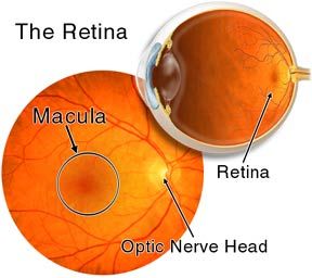
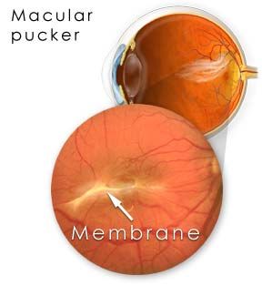
The macula normally lies flat against the back of the eye. An epiretinal membrane (macular pucker) is when wrinkles, creases or bulges form on the macula.
What causes an epiretinal membrane?
As you age, the vitreous--the clear, gel-like substance that fills the middle of your eye begins to shrink and pull away, scar tissue may develop on the macula. Sometimes the scar tissue can shrink and contract, causing the retina to wrinkle or bulge. Eye conditions that are associated with macular pucker include:
- Vitreous detachment (separation of gel inside eye)
- Torn or detached retina
- Inflammation inside eye
- Severe injury to the eye
- Occlusions of the retinal blood vessels
Macular pucker is not usually related to any medical problem outside the eye.
What are the symptoms of macular pucker?
Symptoms of macular pucker range from mild to severe and may involve one or both eyes. Symptoms may include:
- Blurred central(detail) vision
- Distorted, or wavy vision
- Gray and/or cloudy area in central vision
- Central blind spot
- Difficulty reading or performing tasks that require detail vision
How is macular pucker detected?
Your ophthalmologist detects macular pucker by examining your retina. A test called a fluorescein angiogram, a procedure that takes special photographs of the eye may be performed. These photographs show if abnormality exists in your retina. An OCT, a specialized type of ultrasound may also be performed.
How is macular pucker treated?
For mild symptoms, no treatment may be necessary. Updating your eyeglass prescription or wearing bifocals may improve vision. Eye drops, medicines or laser surgery do not improve vision. For more severe symptoms, a surgery called vitrectomy is recommended. The surgery is usually performed as an outpatient procedure in an operating room. During surgery, your retina specialist uses tiny instruments to remove the wrinkled tissue on your macula. After the tissue is gone, the macula flattens and vision slowly improves, though it usually does not return all the way to normal. This is an elective procedure and should be considered if your blurred vision is interfering with your daily activities.
What are the potential risks with vitrectomy surgery?
As with any surgical procedure, rare complications can occur including; infection, bleeding, retinal detachment, recurrence of macular pucker after surgery, cataracts may also develop sooner. Be sure to discuss potential complications with your ophthalmologist before surgery.


Home
......................................
Locations
......................................
Schedule Appointment
About
......................................
Press & Events
......................................
Testimonials
Retina Conditions
......................................
AMD - Macular Degeneration
......................................
Diabetic Eye Disease
......................................
Epiretinal Membrane
......................................
Macular Hole
......................................
Retinal Detachment
Research
......................................
Tools & Resources
......................................
Contact